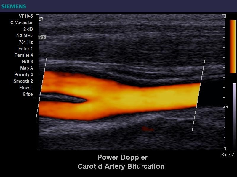

The peak velocity is measured to detect significant stenosis. 1 Pulse-wave Doppler is needed to precisely measure flow velocity, and uses a small sample volume from the vessel center to assess the velocity at that segment. This is called the heel-and-toe technique, and is a method for steering probes. Pushing the head- or foot-side edge creates an angle between the probe surface and the vessel, which can achieve the optimal Doppler angle. 1 In most patients, the probe surface runs parallel to the common carotid artery (CCA) in its usual position, which allows scanning of the carotid artery without applying pressure. However, to accurately record velocity using color Doppler ultrasonography, the angle must be between 30° and 60°. The ultrasound beam should be perpendicular to the skin, with a linear probe generating a grayscale image. Placing a pillow induces a poor evaluation window for the carotid artery and therefore normally should not be used.Īn adequate acoustic angle is important in obtaining an accurate color Doppler image. The neck of the patient should be relaxed and the chin should be slightly raised. Tilting the face too far from the test site can distort the anatomy or compress blood vessels, especially veins. The optimal position for tilting the head of the patient is approximately 45° away from the relevant artery. However, obtaining right-posterior projections is difficult. This position makes it easy to control the ultrasound probe. The examiner generally uses their right hand to evaluate both carotid arteries. A lateral sitting position is used for most ultrasonography examinations. However, the examiner should be familiar with the practice of using both hands. This position provides an expanded sonic window with a clear view of the carotid artery and allows many ultrasound probing positions.


1 In the overhead position, the examiner sits behind the head of the patient beside the end of the bed and uses both hands in the test. You will need to discuss the various treatment options with your doctor in order to find the best approach for you.The examiner can observe the carotid artery from either an overhead or a lateral sitting position. However, if the stenosis is above about 50% or you’re experiencing symptoms, you may need surgery to clear the blockage or hold open the artery. For example, the doctor may recommend a daily dose of aspirin and some changes to your diet. If there is only a small amount of stenosis and you aren’t experiencing symptoms then you may only require lifestyle changes or medication to reduce your stroke risk. The results of your carotid Doppler scan will be explained in detail by your doctor, who will also be able to recommend any treatment that is needed. The best treatment approach will depend on the extent of the blockage and whether you are experiencing any symptoms. If there is any amount of blockage (or stenosis) then you may need treatment. For example, if the doctor tells you that you have 50% stenosis then the blood flow is only half the normal amount. However, if the blood flow through these vessels is impaired, the result will usually be given as a percentage. If no evidence of narrowing is detected in the carotid arteries then your scan results will be reported as normal. You may require treatment if a problem is detected during the carotid Doppler scan in order to reduce this risk and ensure a good blood supply for your brain. If these arteries are narrowed or blocked then it can increase the chances that you will have a stroke.

The carotid Doppler scan is a special type of ultrasound that can be used to check for blockages in the blood vessels that supply your brain.


 0 kommentar(er)
